Extremely close-up images of a cannabis plant, spider eyes and a mouse brain are among the winners of the 50th annual Nikon Small World Photomicrography Competition.
A panel of judges selected 20 winners, shortlisted from around 2,100 entries, for capturing the smallest details in extraordinary clarity.
“Sometimes, we overlook the tiny details of the world around us,” Eric Flem, senior manager of CRM and communications at Nikon Instruments, said in a statement shared with Live Science. “Nikon Small World serves as a reminder to pause, appreciate the power and beauty of the little things, and to cultivate a deeper curiosity to explore and question.”
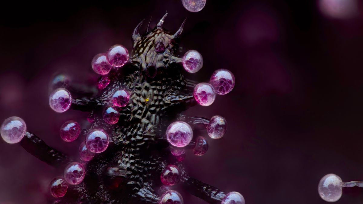
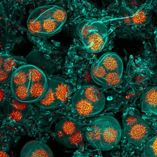
Bruno Cisterna and Eric Vitriol, both researchers at the Department of Neuroscience and Regenerative Medicine at Augusta University in Georgia, won first place with their image of differentiated mouse brain tumor cells — where the cell has developed specialized functions or features. Their image reveals how disruptions in the cell cytoskeleton (which maintains the cell’s shape and enables it to carry out essential functions) can lead to neurodegenerative diseases like Alzheimer’s.
Astronomer-turned-photographer Marcel Clemens was awarded second place for his image of an electrical arc between a pin and a wire, while marijuana photographer Chris Romaine won third place for his image of a cannabis plant leaf.
You can see all 20 winning images below.
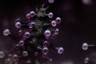
The image of differentiated mouse brain tumor cells taken by Bruno Cisterna and Eric Vitriaol won 1st place in the competition. (Image credit: Dr. Bruno Cisterna & Dr. Eric Vitriol/Nikon Small World 2024)
This image by Marcel Clemens shows an electrical arc between a pin and a wire. (Image credit: Dr. Marcel Clemens/Nikon Small World 2024)
This image, taken by Chris Romaine, shows the leaf of a cannabis plant. The bulbous glands are trichomes — a small hair or growth from the epidermis of a plant — while the purple bubbles contain cannabinoids. (Image credit: Chris Romaine/Nikon Small World 2024)
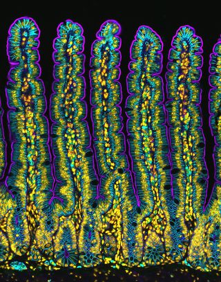
This image, taken by Amy Engevik, shows a section of a small intestine of a mouse. (Image credit: Dr. Amy Engevik/Nikon Small World 2024)
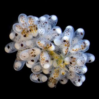
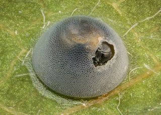
A close-up of a cluster of octopus eggs taken by Thomas Barlow and Connor Gibbons. (Image credit: Thomas Barlow & Connor Gibbons/Nikon Small World 2024)
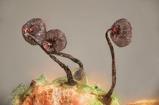
Henri Koskinen’s photograph of the slime mold Cribraria cancellata.(Image credit: Henri Koskinen/Nikon Small World 2024)
A photograph showing the cross section of a leaf from the European beach grass Ammophila arenaria taken by Gerhard Vlcek. (Image credit: Gerhard Vlcek/Nikon Small World 2024)
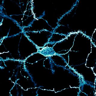
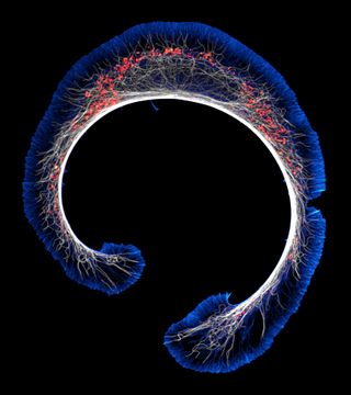
Stephanie Huang’s image shows a neuron from the brain of an adult rat. (Image credit: Stephanie Huang/Nikon Small World 2024)
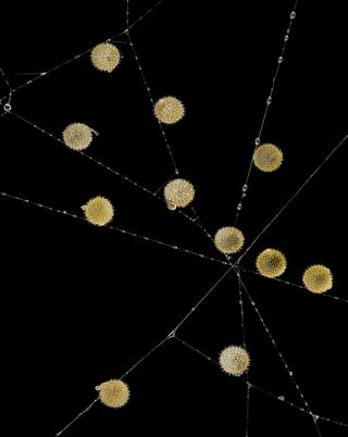
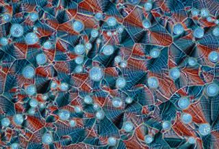
John-Oliver Dum’s photograph shows pollen caught in the web of a garden spider. (Image credit: John-Oliver Dum/Nikon Small World 2024)
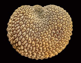
The spores of the black truffle Tuber melanosporum taken by Jan Martinek. (Image credit: Jan Martinek/Nikon Small World 2024)
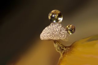
Ferenc Halmos’ image showing water droplets on slime mold on a rotten twig. (Image credit: Dr. Ferenc Halmos/Nikon Small World 2024)
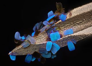
This image, taken by Daniel Knop, shows the wing scales of a butterfly on a medical syringe needle. (Image credit: Daniel Knop/Nikon Small World 2024)
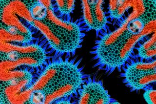
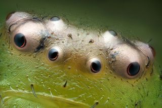
Paweł Błachowicz captured the eyes of the green crap sider Diaea dorsata.(Image credit: Paweł Błachowicz/Nikon Small World 2024)
Marek Miś’ image shows the recrystallized mixture of hydroquinone, a compound that reduces melanin production, and myoinositol, a type of sugar found in the body but also some foods and supplements. (Image credit: Marek Miś/Nikon Small World 2024)
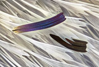
Sébastien Malo’s photograph shows the scales of the Madagascan sunset moth wing Chrysiridia ripheus.(Image credit: Sébastien Malo/Nikon Small World 2024)
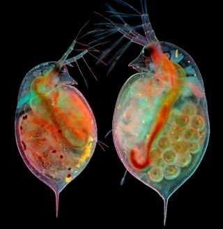
Marek Miś also captured an image of two water fleas with embryos and eggs. (Image credit: Marek Miś/Nikon Small World 2024)
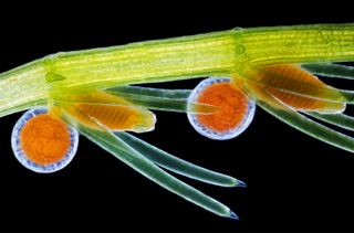
Frantisek Bednar’s image shows the reproductive organs of the stonewort algae Chara virgata. (Image credit: Dr. Frantisek Bednar/Nikon Small World 2024)
An image of an insect egg parasitized by a wasp taken by Alison Pollack. (Image credit: Alison Pollack/Nikon Small World 2024)
Allison Pollack also captured this image of a seed of a silene plant. (Image credit: Alison Pollack/Nikon Small World 2024)
Bruno Cisterna and Eric Vitriol also entered this image of an early stage mouse glioblastoma. (Image credit: Dr. Bruno Cisterna & Dr. Eric Vitriol/Nikon Small World 2024)
Get the world’s most fascinating discoveries delivered straight to your inbox.